DEXcam™ 4 HD Intra-Oral Camera
Visualization that makes a difference in treatment acceptance
Lawrence Spindel, DDS
As an initial user of the DEXcam™ 4 HD (DEXIS, LLC.) for the past three months, I have been working with the camera to report on its technical performance, while keeping a close eye on its clinical efficiency and patient feedback.
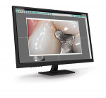
As a clinician, I use 4.4 magnification loupes, so I have no trouble visualizing problems in the mouth. However, when I explain dental issues to my patients and show them their teeth with a little dental or hand mirror, they do not have the benefit of that magnification. With DEXcam 4 HD, I can adjust the magnification so that patients can view their teeth on the screen with much the same clarity as I can with loupes.
When patients see their teeth while hearing my explanation, they can better understand the whole picture. For example, after I open a tooth and remove the filling material, I can use this camera’s live video or captured images to emphasize that the tooth structure is compromised and needs to have a crown to protect it.
In addition to patient education, I also have found that digital photographs are helpful with filing insurance claims. More and more insurance companies seem to be rejecting claims because of lack of information, and these clear, enlarged camera images can help to better illustrate problem areas. For instance, an X-ray of an occlusal surface restoration does not seem very compelling if I am filing a predetermination for a crown. However, if I can include a digital pre-operative CariVu image, intraoral photographs that show the size of the restoration and/or fractures, and a photograph after the restoration is removed, the insurance reviewer can see how little supportive tooth structure remains.
Although some dentists argue that digital cameras do not fit into their budget, it is clear to me that if used properly, the DEXcam 4 HD will pay for itself. When patients can see why a crown is a better clinical choice than a filling in certain circumstances, they are more likely to accept my treatment recommendations.
To conclude, this intraoral camera has many features that are valuable in my workflow. Before I click an image, I can take a video of the area, and when I am ready, I can click again to capture the still photo. In the acquisition software, DEXcapture, I can create a preset for a specific tooth or teeth that I want to capture, and if I want to check all of the teeth, the “click-to-assign” button allows me to assign numbers after the fact. With respect to portability, the quick disconnect feature allows the camera to be moved easily from room to room. And finally, the DEXcam 4 HD integrates flawlessly with my DEXIS software. I can store my radiographs, CariVu images, and digital photographs in the same place and have easy access to any or all of them.
For more information, contact:
DEXIS, LLC
888-553-3947
www.dexis.com
About the Author
Lawrence Spindel, DDS, maintains a private dental practice in Manhattan, New York City, specializing in comprehensive dental care, including cosmetic, implant, and preventive dentistry. He has participated in many clinical trials and is regularly involved with a number of study clubs. Dr. Spindel has published his “Ask Dr. Spindel” dental blog since 2005.